


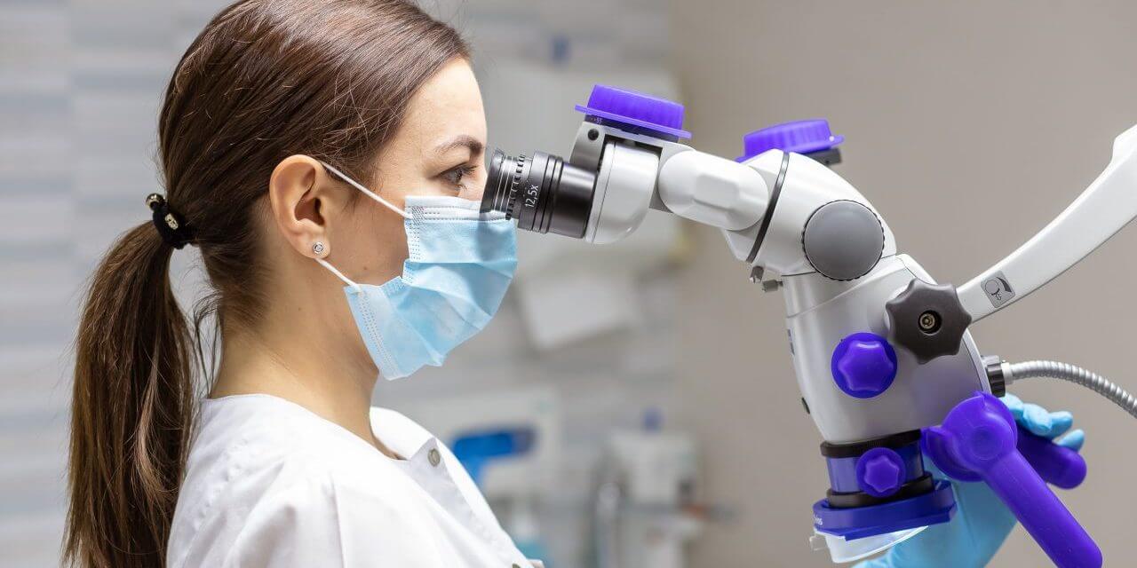
The target image is the most common method of examination of the oral cavity, which is used to identify various diseases and confirm the diagnosis. Special attention is paid to the quality diagnostics in the clinic of Professor Vesova.
The target image of the tooth is done using a radiovisiograph (digital X-ray machine). It gives the opportunity to get images of perfect quality, which are highly informative for the doctor. In addition, it has a low dose of radiation, so this examination is completely safe for health.
Target X-ray imaging makes it possible to identify hidden processes that cannot be detected during a routine examination, confirm the diagnosis and select the optimal method of treatment. Also, such studies are carried out for intermediate control of treatment (restoration, canal filling, etc.).
X-RAY INDICATIONS
- Caries (for diagnosing the degree of damage, including under the filling or prosthesis).
- Endodontic treatment (assessment of roots and canals).
- Granulomas or tooth cyst.
- Defects in the dentition.
- Control treatment.
- Removal of a tooth.
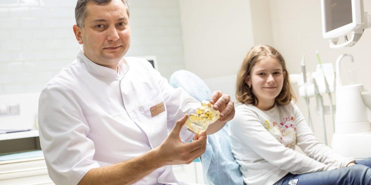
Modern equipment used for targeted X-ray, has a very low level of radiation, so this diagnostic method can be used even during pregnancy (in the 1st and 3rd trimesters with maximum protection). Also, children age is not a contraindication of research.
ADVANTAGES OF RADIOVISIOGRAPHY
- Reduced radiation levels that provide safety for human health.
- High definition of the received images.
- Images are stored in electronic form, so the doctor can quickly find studies of past years if necessary.
- The digital image can be enlarged, which allows the doctor to examine it in the smallest details.
Despite all the advantages of this research technique, targeted X-ray diffraction is not a universal one. The study area is small (1-2 teeth), the picture is taken only in one projection. Therefore, in some cases, a panoramic image or computed tomography is required. Any examination is appointed by our specialists only after the check-up, during which a pre-diagnosis is made.
Price list
Diagnostics

About Us
Welcome to the clinic of dental surgery and dentistry prof. VesovaWe combine art and scientific approach to improve the quality of life of our patients.
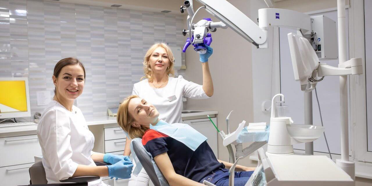
We provide a high level of service and care for the patient, and our efforts are aimed at ensuring that your every visit to our clinic is the most comfortable. A unique synthesis of advanced technologies, an experienced team of specialists and an individual approach give us the opportunity to solve dental problems of any complexity.
Professor Elena Petrovna Vesova
The founder and medical director of the clinic Doctor of Medical Sciences, author of more than 150 scientific articles
OUR ADVANTAGES


Author’s clinic

Transparent price

Collegial approach

Modern equipment

100% safety

Cozy space
Blog

Leave your review about us!
Your opinion is valuable to us